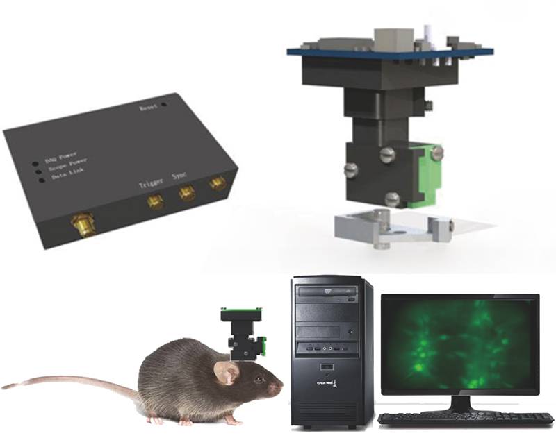
At present, the technologies for studying animal brain related functions mainly include magnetic resonance imaging (fMRI), multi-channel electrophysiology, and two-photon fluorescence microscopy imaging. However, these methods have their shortcomings, such as low spatiotemporal resolution, lack of cell specificity, inability to be used for freely moving animals, high cost, large system, and only suitable for research on head fixation and anesthetized animals.
In response to the limitations of existing technologies, the Qianao Xingke technology research team has developed a super miniature microscopic imaging system that can be used for in vivo calcium imaging of freely moving animals. This system is inexpensive, easy to use, portable, and efficient. The application requirement for recording neural activity calcium signals at the single-cell level on free moving animals has been achieved. Greatly promoting neuroscience research and enriching the technical means of researchers in animal research.
Qianao Xingke provides a variety of ultra micro microscopic imaging systems to meet scientific research needs in multiple directions. The equipment series are as follows:
1、 Ultra Micro Microscopic Imaging System
2、 Ultra Micro Microscopic Imaging System&Automatic Focusing
3、 Ultra Micro Microscopic Imaging System&Photogenetics
4、 Dual color ultra micro microscopy imaging system
5、 Dual color ultra micro microscopy imaging system&autofocus (upcoming)
1、 Ultra Micro Microscopic Imaging System
1. Experimental principle and process
◆ Expressing GCaMP or other calcium ion fluorescent indicators by injecting viruses, implanting GRIN lenses for subsequent observation of changes in cell activity, and waiting for 2-3 weeks for virus expression;
At rest, cells containing GCaMP will exhibit basal fluorescence signals. When cells (astrocytes or neurons) are stimulated, the intracellular calcium level increases, leading to an increase in fluorescence intensity. Fluorescence is collected through implanted lenses, converted into image signals by CMOS, and collected by high-speed image acquisition cards. The change in fluorescence signal intensity can determine (df/f) and reflect the activity of cells;
Image processing software further analyzes the correlation between neural cell activity and behavior.
2. Characteristics
System components include microscope body, fixing plate, GRIN lens, CMOS, image acquisition card and acquisition software, diverter, etc;
Record the calcium signals of group neurons at the single-cell resolution level;
◆ Suitable for in vivo experiments on freely moving animals, recording their behavior in their natural state while observing their brain activity;
By implanting GRIN lenses, deep brain imaging can be achieved;
The system is small in size and lightweight, allowing mice to freely move and conduct behavioral experiments.
3. Usage scenarios
Mainly used for in vivo calcium imaging of behavioral animals, to study the relationship between neuronal circuits and behavior in different parts. Intuitively reflect the activation status of corresponding brain regions under various behavioral paradigms, and conduct real-time observation of deep brain blood vessels and extracellular matrix.
◆ Can complete the following functions:
1. Real time observation of neural projection activity in animals during complex behaviors;
2. Deep brain area calcium ion imaging;
3. Cortical calcium ion imaging;
4. Changes in fluorescence cell migration in deep brain regions.

2、 Ultra Micro Microscopic Imaging System&Automatic Focusing
1. Characteristics
Brand new mirror body design, with a larger field of view;
◆ Updating and upgrading the collection software to better meet scientific research needs;
◆ Paired with video synchronization behavioral software;
◆ Software control for electronic autofocus, achieving clear and accurate imaging of animal brain regions;
◆ Reduce manual focusing interference and ensure more stable data acquisition.
2. Usage scenarios
Wide field fluorescence microscopy is an important means of optical imaging of neuronal activity. Combined with corresponding fluorescent probes, a wide field fluorescence microscope can perform monochromatic and multi-color (such as bicolor, tricolor) fluorescence imaging of neuronal activity.
The automatic focusing ultra micro imaging system is a miniaturized wide field fluorescence microscope that includes micro optical devices, micro imaging elements, and micro mirror structures. It can accurately locate the target area, greatly improving imaging quality, and is an ideal solution for in vivo neural activity optical imaging of free moving animals. Currently, it has been widely applied in neuroscience research both domestically and internationally.
3、 Ultra Micro Microscopic Imaging System&Photogenetics
1. Characteristics
◆ Collecting software updates and upgrades for a better user experience;
◆ The use of external light sources reduces the weight of the mirror body and has a relatively small impact on the activity of experimental animals;
Based on a brand new optical system design, the weight of the mirror body is further reduced and the volume of the mirror body is reduced;
A brand new lighting path design can achieve better fluorescence excitation and spot quality of photoinduced laser, thereby achieving better imaging results;
The external light source end can be freely combined and coupled with different light sources according to different situations, achieving multi-color fluorescence imaging and in situ photogenetic imaging respectively;
◆ Can be equipped with video synchronization behavior software to synchronize/sequentially perform light stimulation and calcium imaging.
2. Main uses
◆ Mainly used to combine in vivo calcium imaging of photogenetic behavioral animals, and further study the relationship between neural circuits and behavior in different parts, intuitively reflecting the activation status of corresponding brain regions under various behavioral paradigms, for real-time observation of deep brain blood vessels and extracellular matrix;
Reduce experimental errors and record at the same position;
◆ Realize cell type specificity.
4、 Dual color ultra micro microscopy imaging system
1. Characteristics
The dual color ultra micro microscopy imaging system adds a 561nm channel on the basis of 470nm excitation, which can simultaneously record green fluorescence and red fluorescence signals at the same site.
When the reference channel uses red fluorescent proteins such as mCherry as markers, the signal of this channel can be used as control data to exclude motion noise (including rotational noise of fiber slip rings) and verify the data validity of the calcium signal channel.
In summary, this system can simultaneously record the activity of two types of neurons in the relevant brain regions in a certain behavioral paradigm, reflecting the encoding characteristics of different types of neurons in the same behavioral paradigm.
2. Application
Mainly used for in vivo calcium imaging of behavioral animals with specific cell types, in order to study the relationship between neuronal circuits and behavior in different parts.
More equipment details and scientific research dry goods can be obtained by following Qianaoxingke's WeChat official account or contacting the regional sales managers